mail_outline sales@mediastorehouse.com
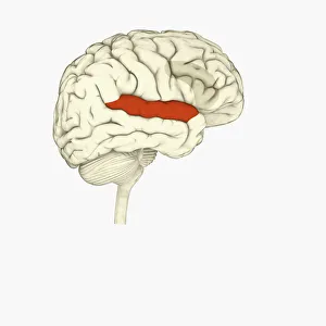
Digital illustration of right superior temporal sulcas, and anterior cingulate cortex highlighted in red and grey in human brain
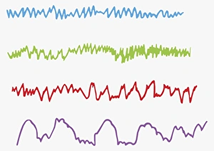
Digital illustration of alpha, beta, theta and delta brain waves
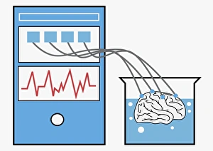
Digital illustration of computer providing stimulation to human brain, and brain experiencing virtual world
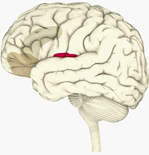
Digital illustration of anterior insular, anterior cingulate cortex, and ventromedial prefrontal cortex highlighted in human brain
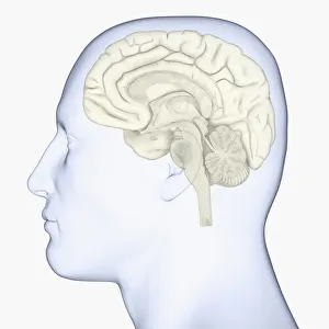
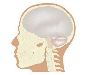
Digital illustration of head in profile showing skull, brain, and spine
Black and white digital illustration of cingulate cortex area of human brain
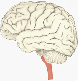
Digital illustration of human brain with spine highlighted in pink
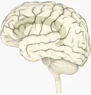
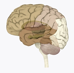
Digital illustration of head in profile showing brain
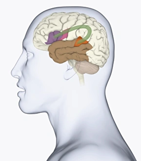
Digital illustration of head in profile showing memory areas of brain
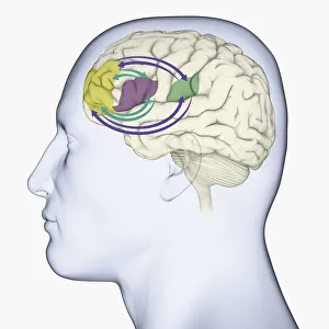
Digital illustration of head in profile showing bundle of nerve fibres connecti ng Brocas area and Wernickes area in human brain
Digital illustration of areas associated with memory in human brain
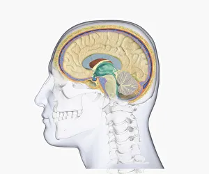
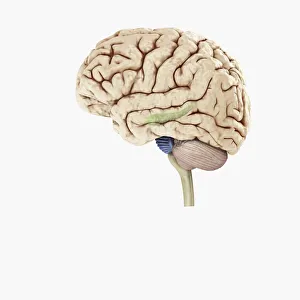
Digital illustration of hippocampus (green) and pons (blue) in left hemisphere of human brain
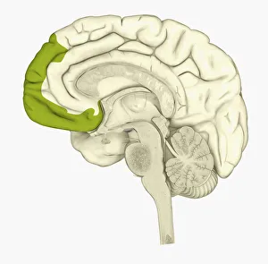
Digital illustration of human brain showing frontal cortex in green
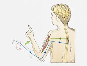
Illustration of reflex action in human arm
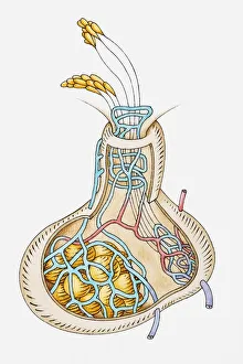
Illustration of pituitary gland releasing hormones
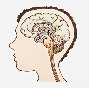
Illustration of human brain
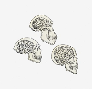
Illustration of skulls of Australopithecus, Homo erectus and Homo sapiens
Cross section biomedical illustration of the brain and skull at 18 years of age
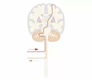
Cross section biomedical illustration of voluntary pathway of human brain
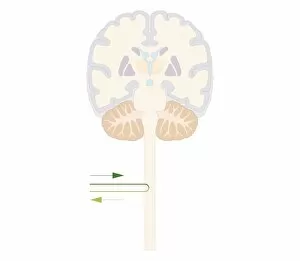
Cross section biomedical illustration of spinal reflex pathway of human brain
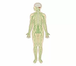
Cross section biomedical illustration of structure of the nervous system in adult male
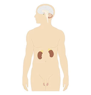
Cross section biomedical illustration of man endocrine system
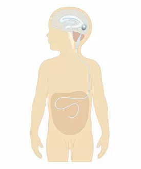
Cross section biomedical illustration of cerebral shunt with valve inserted in brain of boy to remove excess cerebrospinal fluid with tube to carry into stomach
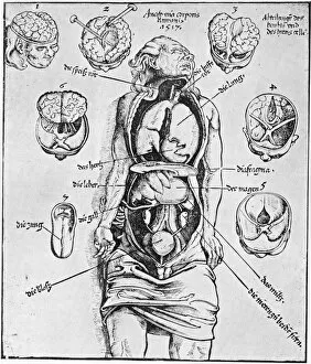
Anatomia Corporis HumaniA diagram of the human brain and other organs. Anatomia Corporis Humani from Spiegl der Arztny by Lorenz Fries, 1518. The diagrams are based on a dissection carried out by German physician Wendelin
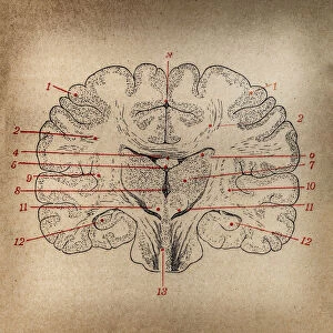
Brain section