mail_outline sales@mediastorehouse.com
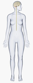
Digital illustration of woman with head in profile showing brain connected to spinal cord
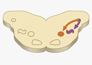
Digital cross section illustration of impulses passing from nearer cochlea nucleus to lateral superior olive
Digital cross section illustration of area signal received in cell of ventral cochlea in human brain
Digital illustration of highlighted areas in human brain affected by motor disorders
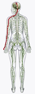
Digital illustration of of human nervous system
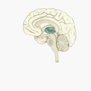
Digital illustration of thalamus in human brain highlighted in green
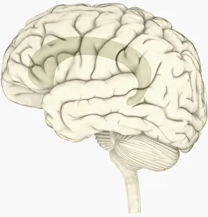
Digital illustration of female human brain
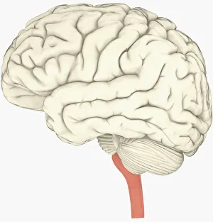
Digital illustration of male human brain
Digital cross section illustration of localization of source of sound in human brain
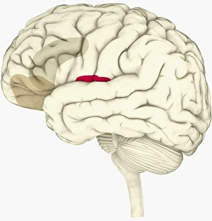
Digital illustration of insula in human brain highlighted in red
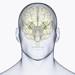
Digital illustration of head in profile showing face recognition area and amygdala in brain
Digital illustration of head in profile showing lateral view of cortex in brain
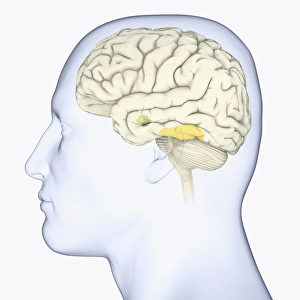
Digital illustration of head showing location of hippocampus in human brain
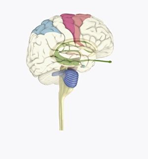
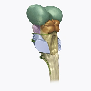
Digital illustration of human brain stem with cerebellum removed revealing medulla and axons
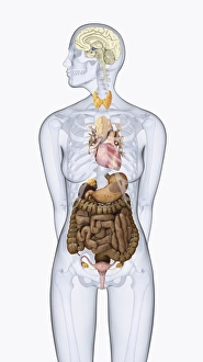
Digital illustration of human neuroendocrine system in female body
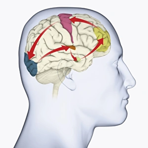
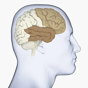
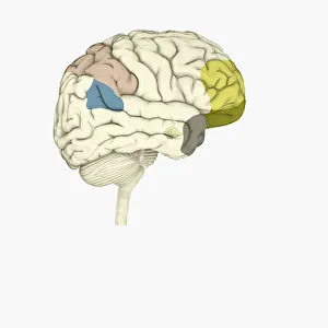
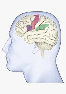
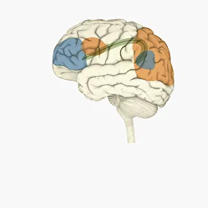
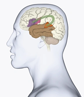
Digital illustration of head in profile showing motor cortex (pink), frontal area and amygdala (green), auditory cortex (orange), and visual cortex in brain
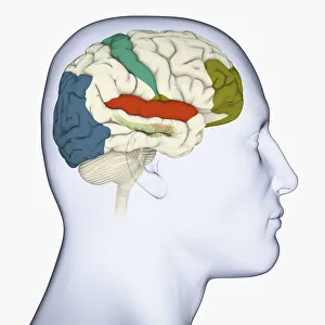
Digital illustration of head in profile showing visual cortex (blue), motor cortex (pink), auditory cortex (orange), frontal area and amygdala (green) brain
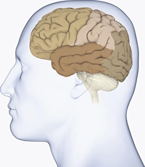
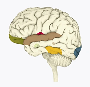
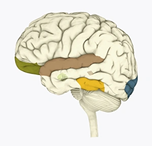
Digital illustration of head in profile showing frontal lobe and temporal lobe in brain
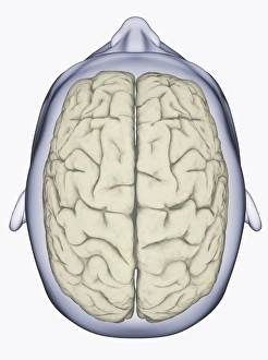
Digital illustration of head showing left and right areas of brain seen from above
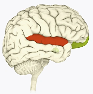
Digital illustration of superior temporal sulcas (red), orbitofrontal cortex and amygdala (green), in human brain
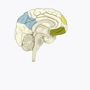
Digital illustration of posterior cingulate cortex (blue), medial frontal gyrus (yellow), and orbitofrontal prefrontal cortex (green) in human brain
Digital illustration of parietal lobe (green), posterior superior temporal sulcas (blue), temperal pole (grey), dorsolateral prefrontal cortex, amygdala
Digital illustration of anterior cingulate cortex (grey), and medial frontal cortex (green) in human brain
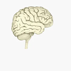
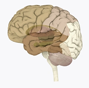
Digital illustration of human brain
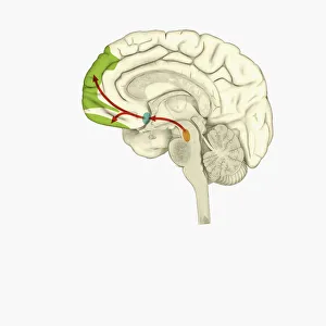
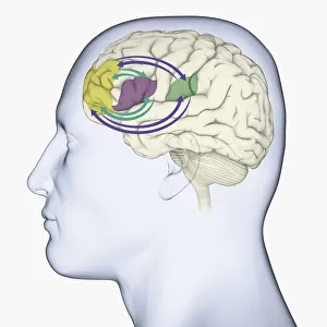
Digital illustration of reward pathway in human brain
Digital illustration of human brain associated with full awareness
Digital illustration of areas of information highlighted in human brain
Digital illustration of human brain with orbitofrontal cortex and amygdala highlighted in green
Digital illustration of head in profile showing mirror neurons in human brain highlighted in purple, pink and green
Digital illustration of head in profile showing area of mirror neuron in brain highlighted in orange
Digital illustration of frontal lobe and parietal lobe areas (orange) in left hemispheres (blue), and pathway of data from parietal lobe to frontal lobe (green) in human brain
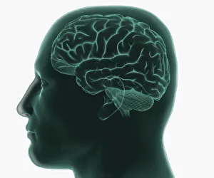
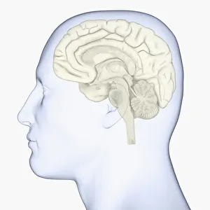
Digital illustration of human head in profile showing brain
Digital illustration of right superior temporal sulcas, and anterior cingulate cortex highlighted in red and grey in human brain
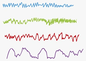
Digital illustration of alpha, beta, theta and delta brain waves
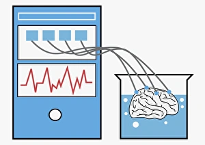
Digital illustration of computer providing stimulation to human brain, and brain experiencing virtual world
Digital illustration of anterior insular, anterior cingulate cortex, and ventromedial prefrontal cortex highlighted in human brain
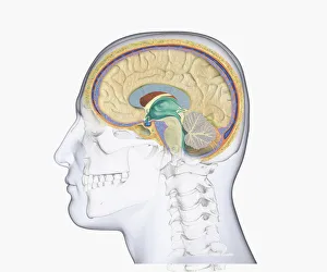
Digital illustration of head in profile showing skull, brain, and spine
Black and white digital illustration of cingulate cortex area of human brain
Digital illustration of human brain with spine highlighted in pink
Digital illustration of head in profile showing brain
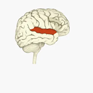
Digital illustration of head in profile showing memory areas of brain
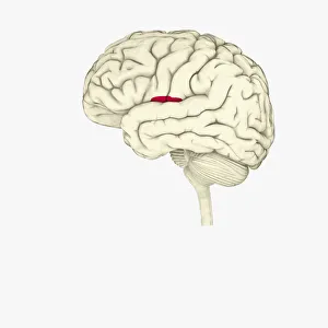
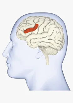
Digital illustration of head in profile showing bundle of nerve fibres connecti ng Brocas area and Wernickes area in human brain
Digital illustration of areas associated with memory in human brain
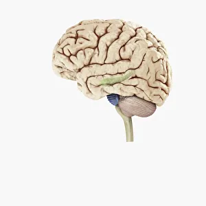
Digital illustration of hippocampus (green) and pons (blue) in left hemisphere of human brain
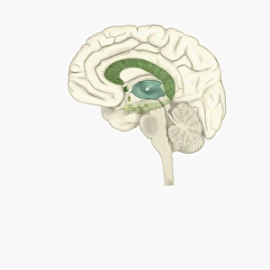
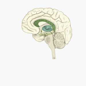
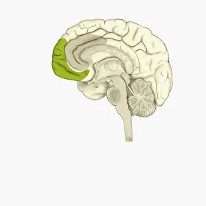
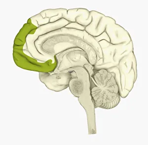
Digital illustration of human brain showing frontal cortex in green
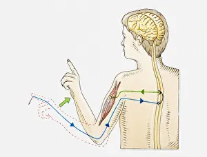
Illustration of reflex action in human arm
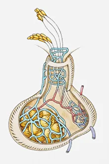
Illustration of pituitary gland releasing hormones
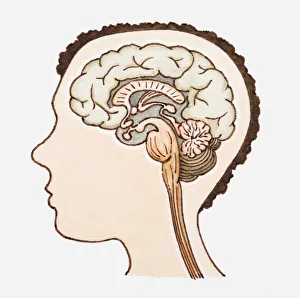
Illustration of human brain