mail_outline sales@mediastorehouse.com
1,282 Photographic Prints
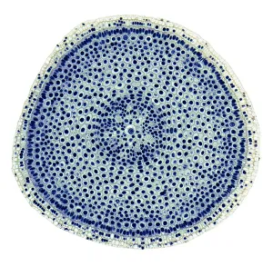
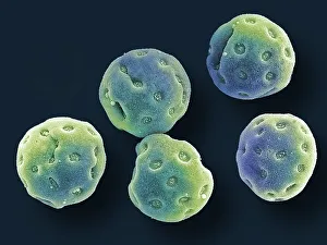
Onion root tip, LMMitosis. Light micrograph (LM) of a transverse section of onion (Allium cepa) root tip to show cells undergoing mitosis (nuclear division). Magnification: x100 when printed at 10 centimetres wide
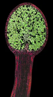
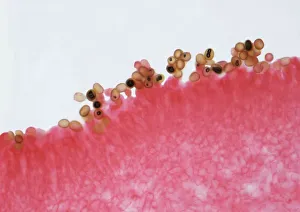
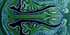
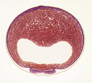
Liverwort spore capsule, LMLiverwort spore capsule. Light micrograph (LM). Longitudinal section through the thallus and sporangium of a liverwort (Pellia epiphylla)
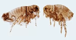
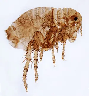
Human fleas, LMHuman fleas. Light micrograph (LM) of a male (left) and female human flea (Pulex irratans). Fleas are wingless and flattened from side to side, which makes them difficult to dislodge in hair
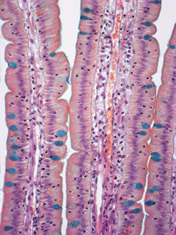
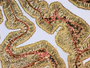
Small intestine, LMSmall intestine. Light micrograph (LM) of a section through the finger-like projections (villi) of the duodenum, the uppermost part of the small intestine
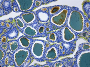
Thyroid, LMThyroid gland. Light micrograph (LM) of a thyroid gland showing the follicles. The follicles are lined by a single layer of cuboidal epithelial cells (blue)
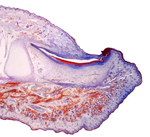
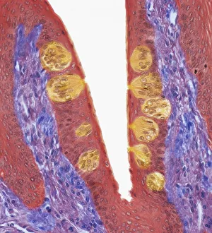
Fingertip, LMFingertip. Light micrograph (LM) of a section through the fingertip. The nail (orange) is at top center, with the nail root below. The nail bed is dark purple and is continous with the epithelium
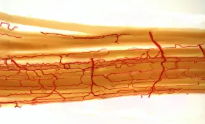
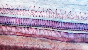
Blood supply to muscles, LMBlood supply to muscles. Light micrograph (LM) showing blood supply to muscle fibers. The muscle fibers (yellow) have been teased apart to reveal the capillary bed (red)
Fallopia tube, LMFallopian tube. Light micrograph (LM).The fallopian tube, or oviduct, conveys the egg from the ovary to the uterus. Ciliated columnar epithelium is yellow
Mushroom gill, LMMushroom gills. High power light micrograph (LM) of a section through the gills of a mushroom, Agaricus sp. (formerly Psalliota sp.)
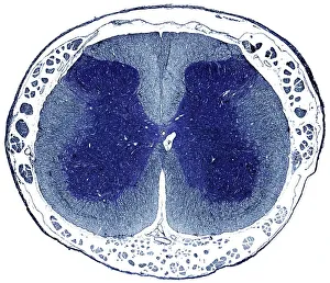
Spinal cord, LMSpinal cord. Light micrograph (LM) of a cross-section through the human spinal cord in the lumbar region. The spinal cord consists of a butterfly-shaped core (dark blue) known as grey matter
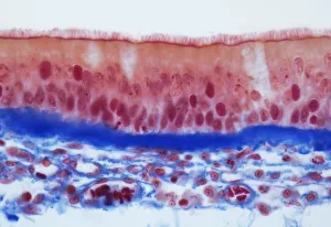
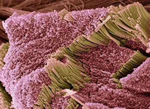
Trachel epithelium, LMTrachea epithelium. Light micrograph (LM) of a vertical section through the pseudostratified columnar epithelium from the trachea
Female flea, LMHuman flea. Light micrograph (LM) of a female human flea (Pulex irratans). Fleas are wingless and flattened from side to side, which makes them difficult to dislodge in hair
Nasal sinuses, LMNasal sinuses. Light micrograph (LM) of the nasal sinuses ( lined by cyan epithelium ) and the supporting cartilages (green). Bone tissue is identified by the blue bone marrow
Xylem, LMXylem tissue. Light micrograph (LM) of a section through sunflower(helianthus annuus) tissue showing spiral tracheids, a type of xylem
Taste buds, LMTaste buds. Coloured light micrograph of a section through the tongue, showing taste buds (round, yellow). The taste buds are within papillae (projections) located on the surface of the tongue
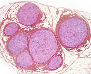
Nerve fibres, LMNerve fibres. Light micrograph (LM) of a transverse section through a bundle (fascicle) of nerve fibres. Within each fascicle are many myelinated nerve fibres
Developing frog egg, LMDeveloping frog egg. Light micrograph of a transverse section through a developing egg laid by a common frog (Rana temporaria). The egg has reached the early neurula stage
Macrophage, SEMMacrophage. Coloured scanning electron micrograph (SEM) of a macrophage white blood cell. Macrophages are cells of the bodys immune system
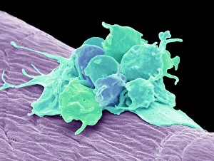
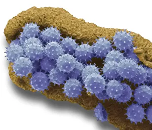
Activated platelets, SEMActivated platelets. Coloured scanning electron micrograph (SEM) of activated platelets attached to surgical gauze. Platelets are tiny blood cells that help the body form clots to stop bleeding
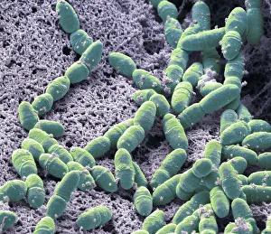
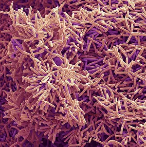
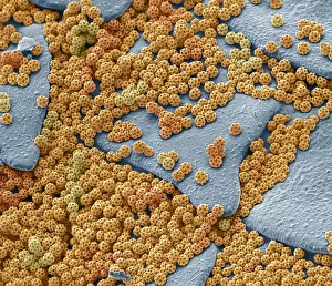
Streptococcus mutans, SEMStreptococcus mutans. Coloured scanning electron micrograph (SEM). S. mutans is a coccoid shaped, Gram-positive, anaerobic bacteria that is part of the normal bacteria flora of the mouth
Plaque-forming bacteria, SEMPlaque-forming bacteria, coloured scanning electron micrograph (SEM). Plaque consists of a film of bacteria embedded in a glycoprotein matrix
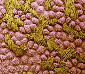
Fallopian tube, SEMFallopian tube. Coloured scanning electron micrograph (SEM) of the surface of a human fallopian tube. Fallopian tubes are ducts that lead from the ovaries to the uterus
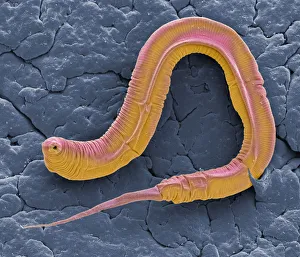
C elegans, SEMCaenorhabditis elegans worm, coloured scanning electron micrograph (SEM). C. elegans is a soil-dwelling hermaphrodite nematode worm and one of the most studied animals in biological
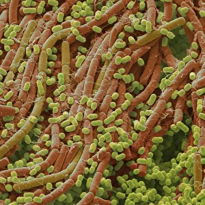
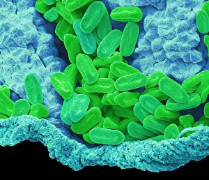
Soil bacteria, SEMSoil bacteria. Coloured scanning electron micrograph (SEM). Bacteria in the soil are directly tied to nutrient recycling especially carbon, nitrogen, phosphorus and sulfur
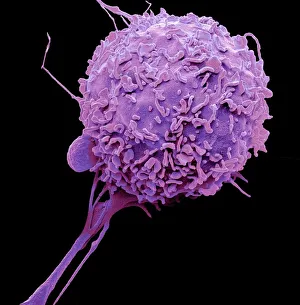
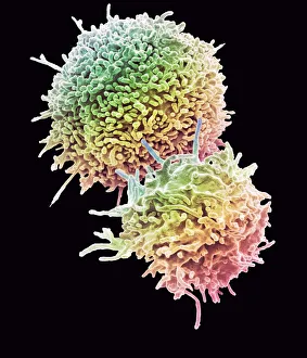
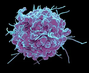
Resting T lymphocytes. Coloured scanning electron micrograph (SEM) of resting T lymphocytes from a human blood sample. T lymphocytes, or T cells
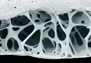
Bird bone tissue, SEMBird bone tissue. Coloured scanning electron micrograph (SEM) of cancellous (spongy) bone from a starlings (Sturnus vulgaris) skull
Kefir bacteria, SEMKefir bacteria. Scanning electron micrograph (SEM) of Lactococcus bacteria from kefir, a fermented milk beverage containing beneficial yeast as well as probiotic bacteria
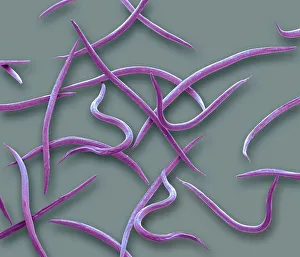
Nematodes, SEMPhasmarhabditis hermaphrodita. Coloured scanning electron micrograph (SEM) of Phasmarhabditis hermaphrodita. Phasmarhabditis hermaphrodita is a microscopic nematode in the family Rhabditidae
Tendon, SEMTendon, coloured scanning electron micrograph (SEM), showing bundles of collagen fibres. The parallel alignment of the fibres make tendons inelastic but flexible. Tendons attach muscle to bone
Brochosomes, SEMBrochosomes, coloured scanning electron micrograph (SEM). Brochosomes are intricately structured microscopic granules secreted by leafhoppers
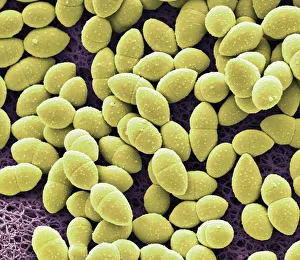
Verbena pollen, SEMVerbena pollen. Coloured scanning electron micrograph (SEM) of pollen grains from verbena bonariensis. Verbena bonariensis is a tall, slender-stemmed perennial
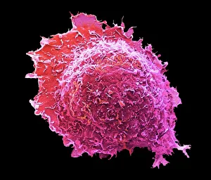
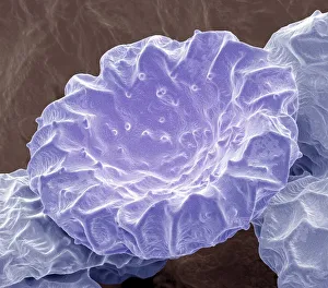
Apoptosis, SEMApoptosis. Coloured scanning electron micrograph (SEM) of a 293T cell in the early stages of programmed cell death, or apoptosis. Apoptosis occurs when a cell becomes old or damaged
Diatoms, SEMDiatoms. Coloured scanning electron micrograph (SEM) of fresh water centric diatom frustules (skeleton), Diatoms are a type of algae (Chromophyta, Bacillariophyceae)
Convolvulus pollen grains, SEMConvolvulus pollen grains. Coloured scanning electron micrograph (SEM) of pollen grains from a convolvulus flower. Convolvulus is a genus of about 200 to 250 species of flowering plants in
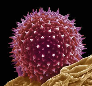
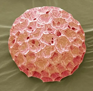
Hollyhock pollen grain, SEMHollyhock pollen grain. Coloured scanning electron micrograph (SEM) of a pollen grain from a hibiscus (Alcea setosa) flower. Pollen grains are the male sex cells of a flowering plant
Osteocyte, SEMOsteoblast bone cell. Coloured scanning electron micrograph (SEM) of an osteoblast bone cell. Osteocytes are osteoblasts (bone-producing cells) that have become trapped within bone cavities (lacunae)
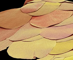
Silverfish scales, SEMSilverfish scales (Lepisma saccharina). Coloured scanning electron micrograph (SEM) of scales from a silverfish. The silverfish is a primitive insect that has remained unchanged for millions of
Water lily pollen grains, SEMWater lily pollen grains. Coloured scanning electron micrograph (SEM) of pollen grains from a water lily flower. Nymphaeaceae is a family of flowering plants, commonly called water lilies
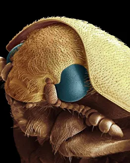
Dermestid beetle, SEMDermestid beetle. Scanning electron micrograph (SEM(=) of a dermestid beetle. Dermestidae are a family of Coleoptera that are commonly referred to as skin beetles
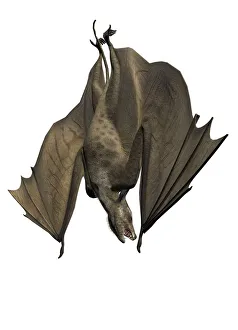
Icaronycteris, illustrationIcaronycteris against a white background, illustration
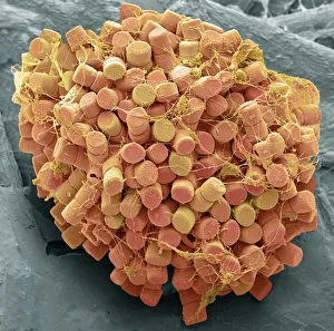
Hibiscus pollen grains, SEMChinese hibiscus pollen. Coloured scanning electron micrograph (SEM) of pollen grains on the anther of a Chinese hibiscus (Hibiscus rosa-sinensis) flower
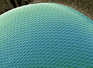
Wasp eye, SEMWasp eye. Coloured scanning electron micrograph (SEM) of numerous lenses, called ommatidia, that make up the compound eye of a wasp (Suborder Apocrita)
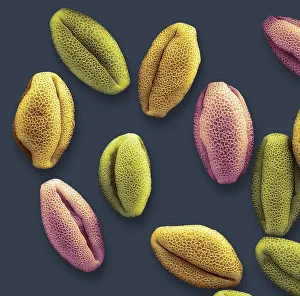
Phlox pollen, SEMPhlox pollen grain, magnified x2000 when printed at 10 centimetres wide
Bougainvillea pollen, SEMBougainvillea pollen grains, magnified x2000 when printed at 10 centimetres wide
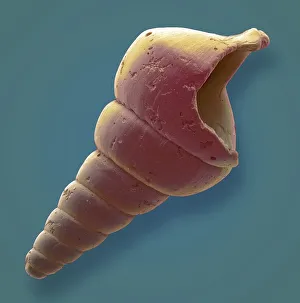
Gastropod microfossil, SEMGastropod. Coloured scanning electron micrograph (SEM) of a gastropod microfossil from maldives beach sand. Microfossils are roughly 0.05 to 2mm in size
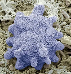
Korotnevella amoeba, SEMKorotnevella. Coloured scanning electron micrograph (SEM) of the scales covering a naked amoeba of the genus Korotnevella
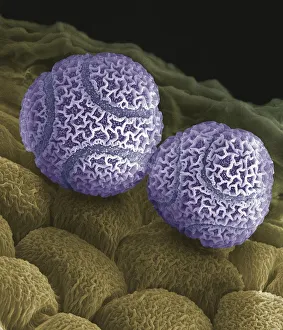
Bellflower pollen, SEMBellflower pollen. Coloured scanning electron micrograph (SEM) of pollen grains from a bellflower (Campanula sp.). Pollen grains are the male gametes (sex cells) of a plant
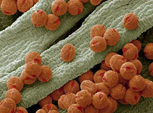
Passion flower pollen. Coloured scanning electron micrograph (SEM) of pollen grains from a passion flower (Passiflora caerulea). Pollen grains are the male gametes (sex cells) of a plant