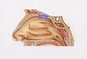
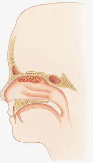
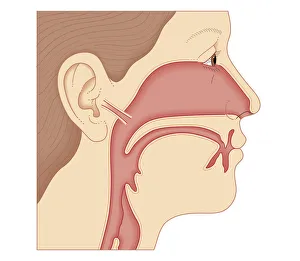
Cross-section illustration of nasal cavity, nasal epithelium, and smell receptors (Olfaction)
Anatomy, Biomedical Illustration, Bone, Close-Up, Connection, Fragility, Human Hair, Human Head, Human Representation, Human Nose, Illustration and Painting, Illustrative Technique, Inside Of
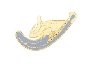
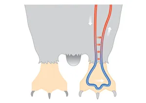
Illustration showing direction air passes through bony crest on top of Parasaurolophus skull
Air, Anatomy, Animal Bone, Animal Themes, Arrow Sign, Bony, Close-Up, Crest, Cross Section, Direction, Extinct, Illustration and Painting, No People, On Top Of, Paleontology, Parasaurolophus
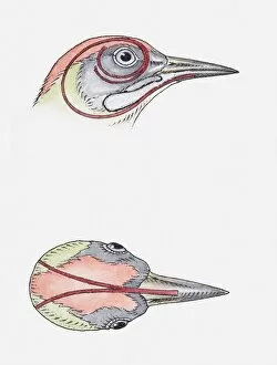
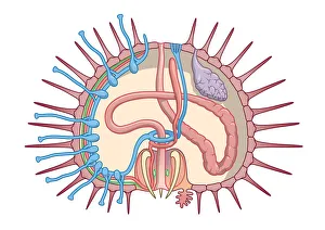
animal head, animal themes, bird, cross section, long, no people, physiology, profile, studio shot, tongue, two animals, vertical, watercolour painting, white background, wildlife, woodpecker