mail_outline sales@mediastorehouse.com
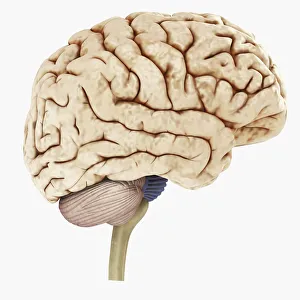
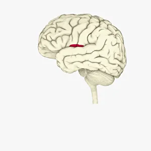
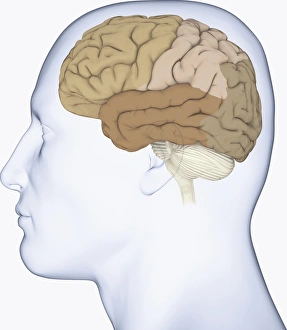
Digital illustration of showing right side human brain
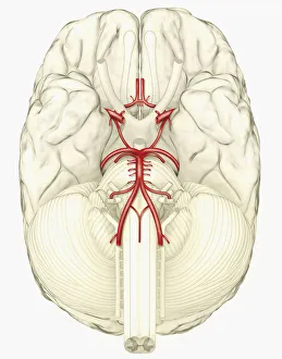
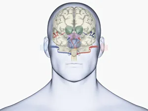
Digital illustration showing arteries in human brain
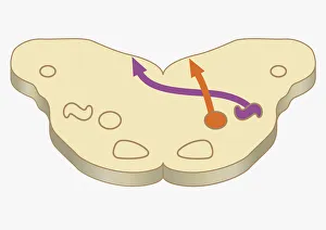
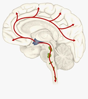
Digital cross section illustration of impulses from lower brain stem passing to inferior colliculus of midbrain
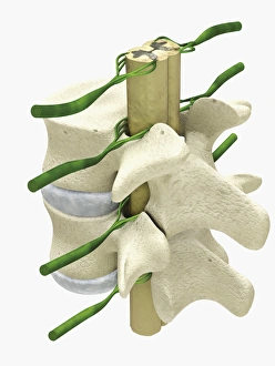
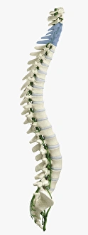
Cross section digital illustration of spinal cord and nerves surrounded by vertebra
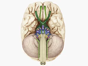
Digital illustration of cranial nerves linked to human brain
Digital illustration of human spine with damaged area highlighted in blue resulting in quadriplegia
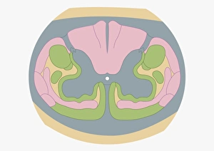
Cross section digital illustration of human brain showing caudate nucleus, putamen, external globus pallidus, and internal globus pallidus
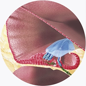
Digital illustration of Corti organ found in cochlea of human ear
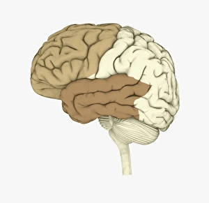
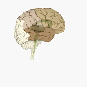
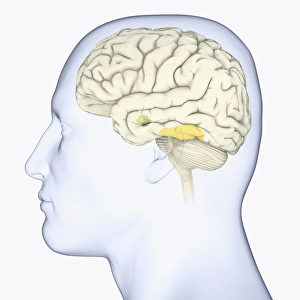
Digital illustration of human brain highlighting frontal and temporal lobes
Cross section digital illustration of spinal nerve fibres and convey motor signals highlighted in pink and green
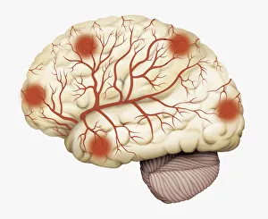
Digital illustration of human brain showing blood vessels, and areas of dead tissue highlighted in red
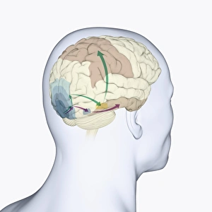
Digital illustration of head in profile showing dorsal and ventral pathways of brain
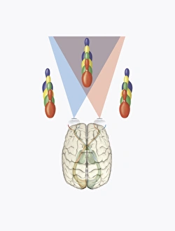
Digital illustration of human brain with differing views provided by each eye producing three dimensional vision
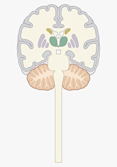
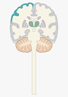
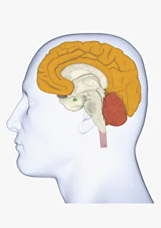
Cross section digital illustration of brain highlighting cerebral cortex, thalamus, and brain stem
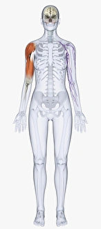
Digital illustration of human skeleton showing upper arm muscles
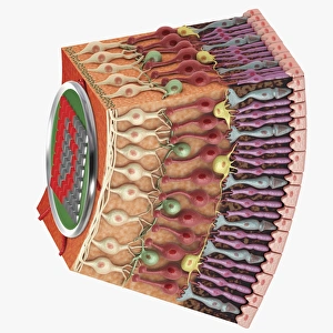
Cross section digital illustration of retina with futuristic retinal implant
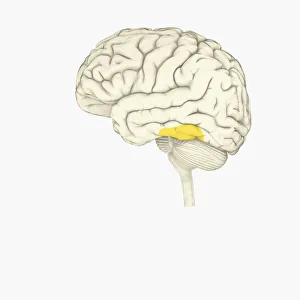
Digital illustration of human brain showing face-recognition area highlighted in yellow
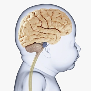
Digital illustration of head of baby in profile showing brain
Digital illustration head in profile with cerebrum and cortex highlighted
Digital illustration of human brain with sound entering via brain stem and thalamus to auditory cortex
Digital illustration of domestic cat showing cerebellum highlighted in orange
Digital cross section illustration of medial nucleus and lateral superior olive in human brain
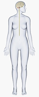
Digital illustration of woman with head in profile showing brain connected to spinal cord
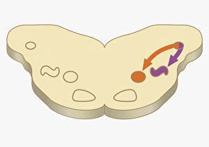
Digital cross section illustration of impulses passing from nearer cochlea nucleus to lateral superior olive
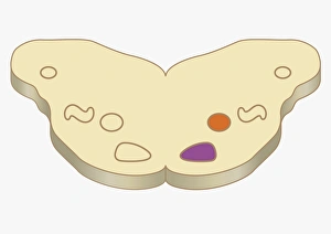
Digital cross section illustration of area signal received in cell of ventral cochlea in human brain
Digital illustration of Hypocretin System in human brain with Hypothalamus highlighted in blue, Locus Coeruleus and Raphne nuclei in green
Digital illustration of highlighted areas in human brain affected by motor disorders
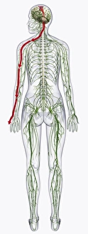
Digital illustration of of human nervous system
Digital illustration of brain areas involved in altered states
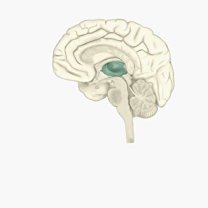
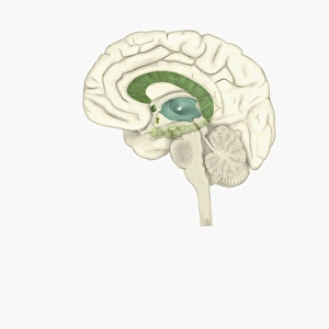
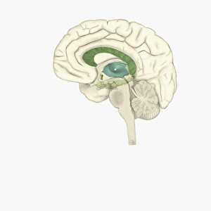
Digital illustration of thalamus in human brain highlighted in green
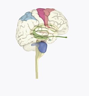
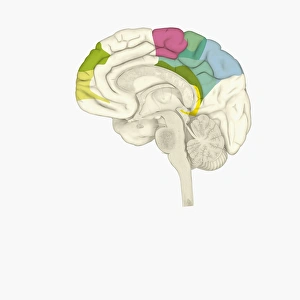
Digital illustration of parts of human brain highlighted in shades of green, pink, blue, and yellow
Digital illustration of female human brain
Digital illustration of male human brain
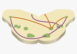
Digital cross section illustration of localization of source of sound in human brain
Digital illustration of insula in human brain highlighted in red
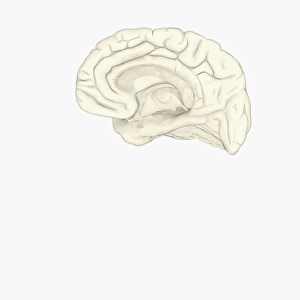
Digital illustration of human brain showing corpus collosum and cingulate gyrus on medial surface of human brain
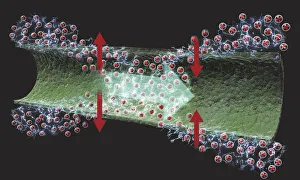
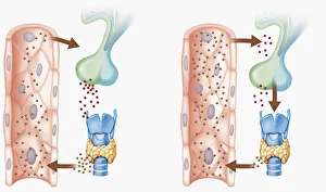
Digital illustration of nerve signal moving along membrane as wave of depolarization and repolarization
Digital illustration of head in profile showing face recognition area and amygdala in brain
Digital illustration of head in profile showing lateral view of cortex in brain
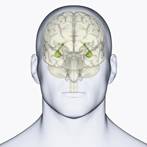
Digital illustration of head showing location of hippocampus in human brain
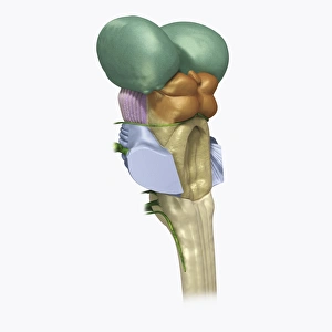
Digital illustration of human brain stem with cerebellum removed revealing medulla and axons
Digital cross section illustration of hypothalamus triggering chain reaction of reduced hormone production resulting in falling glucose levels
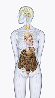
Digital illustration of human neuroendocrine system in female body
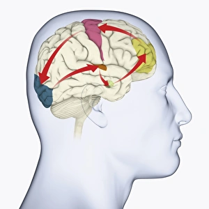
Digital illustration of head in profile showing motor cortex (pink), frontal area and amygdala (green), auditory cortex (orange), and visual cortex in brain
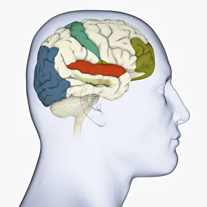
Digital illustration of head in profile showing visual cortex (blue), motor cortex (pink), auditory cortex (orange), frontal area and amygdala (green) brain
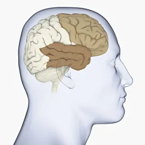
Digital illustration of head in profile showing frontal lobe and temporal lobe in brain
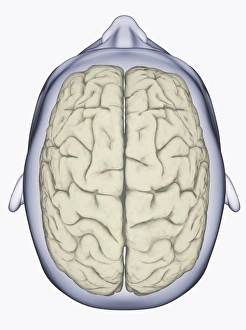
Digital illustration of head showing left and right areas of brain seen from above
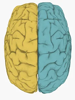
Digital illustration of left hemisphere (yellow) and right hemisphere (blue) of human brain