mail_outline sales@mediastorehouse.com
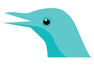
Digital illustration bird head showing straight, insect-eating beak
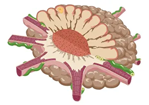
Digital illustration of coral polyp showing nutrients diffused through gastrodermis into tissue, and single digestive opening used as mouth and anus
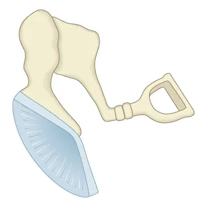
Digital illustration of mammal middle ear showing malleus, incus, and stapes
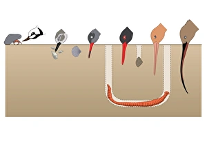
Digital illustration showing feeding strategies of birds and beak their shapes
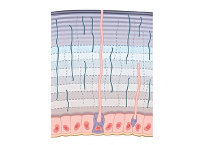
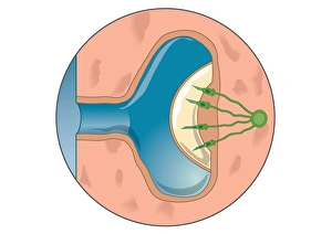
Digital illustration of contracted chromatophore showing densely packed pigment, and nerves controlling muscle fibres which alter pigment dispersal
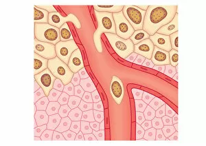
Digital illustration showing secondary tumour where cancerous cells have divided and lodged in narrow blood vessel and surrounding tissue
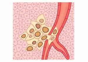
Digital illustration of breached lymph vessel as primary tumour grows and its cells invade adjacent tissues
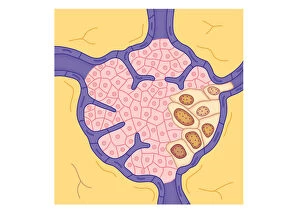
Digital illustration of tumour in lymph node
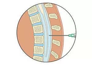
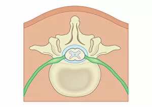
Digital illustration of lumbar puncture using spinal needle inserted into lumbar vertebrae and dura mater
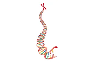
Digital illustration of animal DNA and chromosome
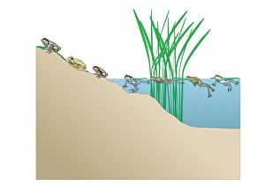
Digital illustration of frogs each having distinctive calls known as dominant frequencies made from different areas of pond
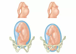
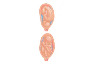
Digital illustration of pregnant two women and position of each babys head entering and inside pelvic area
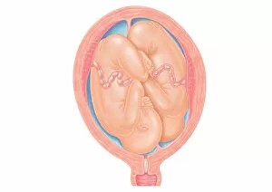
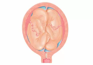
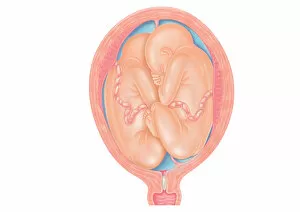
Cross section digital illustration of twins in normal position in uterus
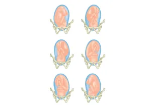
Six cross section digital illustrations of foetus showing position head in pelvis
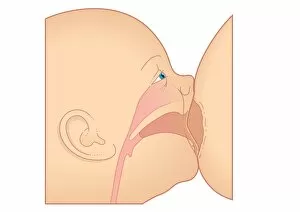
Digital illustration showing baby breastfeeding
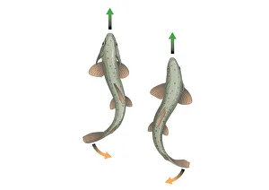
Digital illustration showing body propulsion of fish using tail and fin to generate sideways and backwards thrust in water enabling it to swim in a straight line
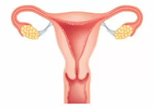
Digital cross section illustration of human uterus, fallopian tubes, ovaries, cervix, and vagina
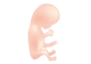
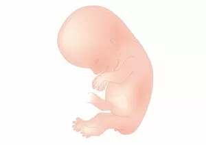
Digital illustration of foetal size at 11 weeks
Cross section digital illustration showing normal and breech positions
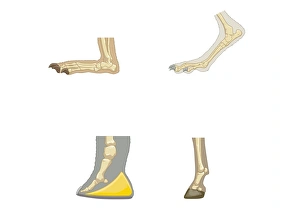
Digital illustrations of showing, plantigrade, digitigrade, and unguligrade walking gaits
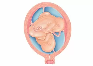
Cross section digital illustration of twins showing normal foetal presentation and transverse lie
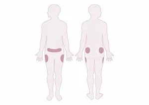
Digital illustration representing areas of fat distribution on belly, hips and thighs
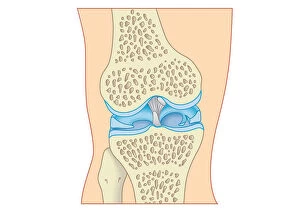
Digital cross section illustration of torn cartilage in knee
Digital cross section illustration of mammalian ear including pinna, ear drum, middle ear
Digital cross section illustration of procuticle divided into hardended exocuticle with many compacted fibres, more flexible endocuticle
Cross section digital illustration of foetus in normal position and breech position
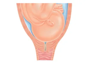
Cross section digital illustration of head of foetus in pelvis, pushing against cervix as labour nears, also showing mucus plug in cervix
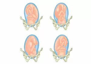
Four cross section digital illustrations showing head of foetus in pelvis
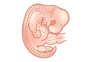
Digital illustration of foetus size at 7 weeks
Digital illustration of foetal size at 10 weeks
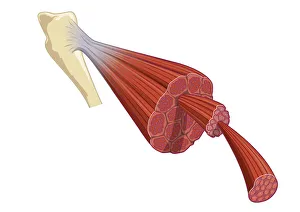
Digital illustration of vertebrate muscle attached to skeleton, consisting of bundles of muscle fibres composed of thousands of thinner muscle fibrils
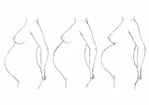
Digital illustration of bump position during pregnancy
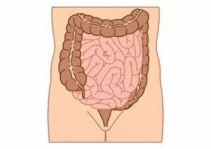
Digital illustration of location of intussusception in small intestine
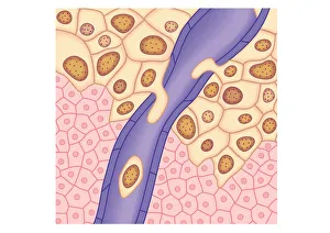
Digital illustration of blood vessel wall rupturing, primary tumour expanding, cells rupturing walls of blood vessels, enabling cancerous cells to detach and spread via blood flow
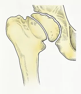
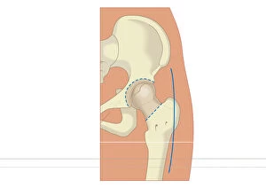
Digital illustration showing degeneration of hip joint known as Perthes disease
Digital illustration Spina Bifida, Meningocele, where protective covering around spinal cord protrudes through malformed vertebra to form sac filled with cerebrospinal fluid
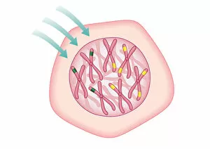
Digital illustration showing temporary cell damage to genes on chromosomes caused by an attacking carcinogen
Digital illustration of prosthetic hip joint replacement
Cross section digital illustration of twins in breech position with buttocks presented first
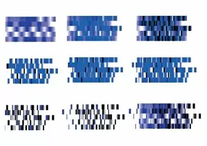
Digital illustration DNA map
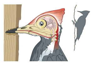
Digital illustration showing woodpeckers shock absorbers connecting chisel shaped beak to skull by
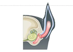
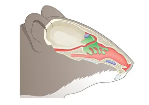
Digital cross section illustration of olfactory system of House Mouse (Mus musculus)
Digital illustration of lateral line system visible as faint line running down each side of fish bod
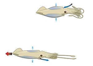
Digital illustration of squid drawing water into mantle cavity using muscles to expand and contract
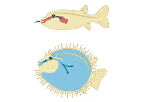
Digital cross section illustration of Porcupine Fish and Puffer Fish abdomen
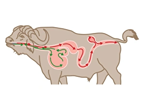
Digital cross section illustration of four chambers in stomach of ruminant, showing rumen, reticulum
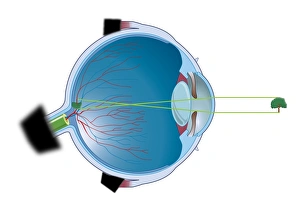
Digital illustration of vertebrate eye using lens to focus light and form and image
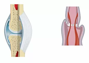
Digital illustration of invertebrate joint, left, and vertebrate joint, right