Physiology Collection (page 7)
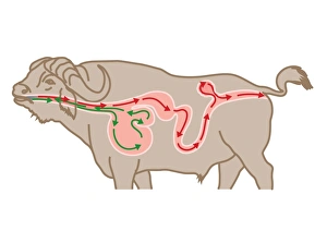
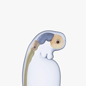
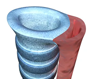
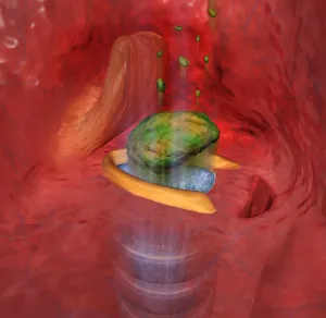
Digital cross section illustration of four chambers in stomach of ruminant, showing rumen, reticulum
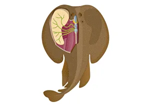
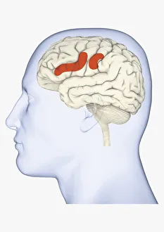
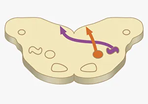
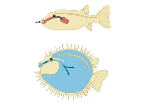
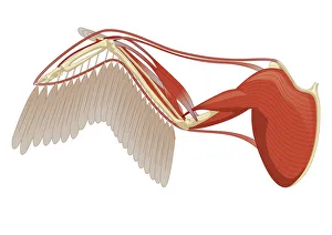
Digital illustration of Electric Ray showing electricity and magnetism in cerebellum, lobus electric
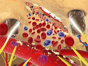
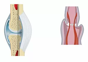
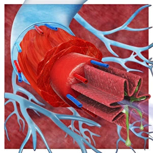
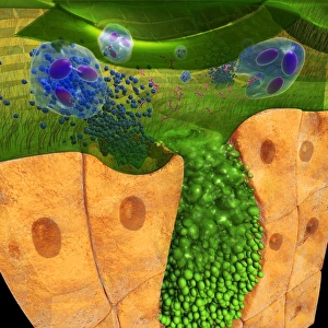
Digital cross section illustration wound below human skin showing red and white blood cells and yell
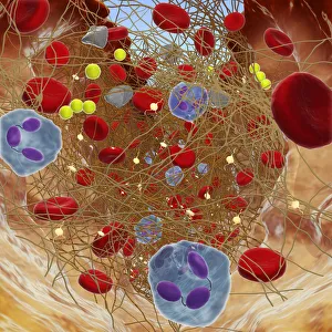
Cross section showing wound below skin, long fibrin threads trapping red blood, yellow platelets cau
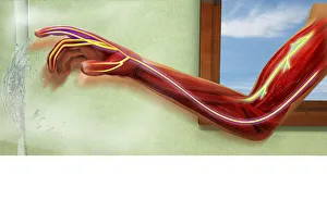
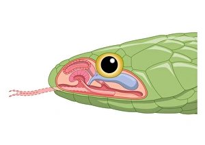
Digital illustration showing sensory neurons sending message to nerves in human arm after scalding b
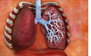
Digital cross section illustration of human lung with pollen allergens in trachea and bronchioles ca
Digital cross section illustration of ciliate cell showing rhinovirus and antobodies in nasal cavity
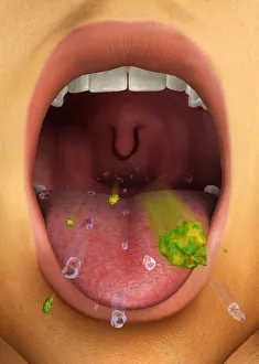

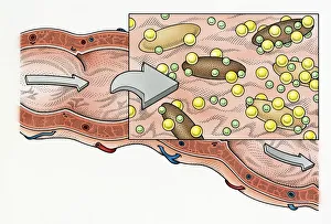
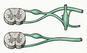
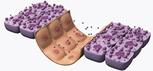
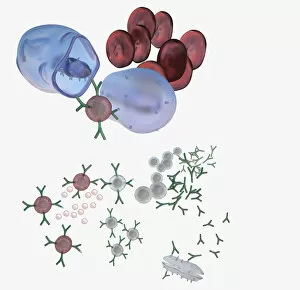
Illustration of immune response, involving chain of defensive white blood cells, triggered by microb
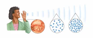
Illustrations of woman shouting showing sound waves and insets of vocal cords, rarefaction and compr