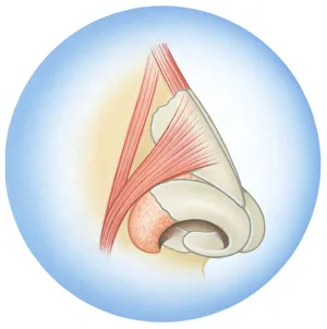
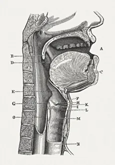
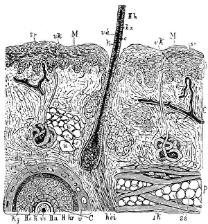
Human speech organs, wood engraving, published in 1880
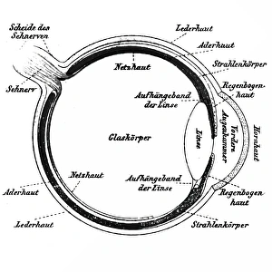
The Organs of Speech: A) mouth, B) uvula, C) tongue, D) tonsils, E) epiglottis, F) thyroid cartilage, G) arytenoid muscle, H+I) vestibular folds, K) vocal fold, L) larynx, M) hypopharynx, N) trachea
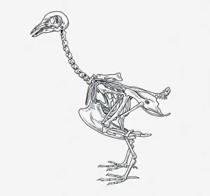
anatomy, animal themes, bird, coracoid, cranium, cut out, femur, humerous, mandible, no people, ostrich, pelvis girdle, pen and ink, phalanges, physiology, profile, pygostyle, side view, skeleton
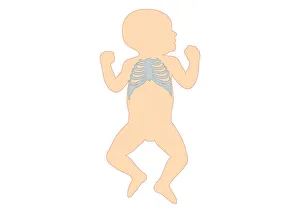
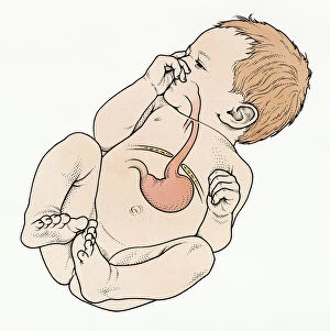
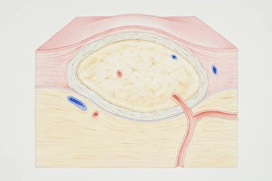
Anatomy, Baby, Beginnings, Biomedical Illustration, Caucasian Appearance, Childhood, Close-Up, Connection, Cross Section, Diaphragm, Digitally Generated, Healthcare and Medicine, Hernia